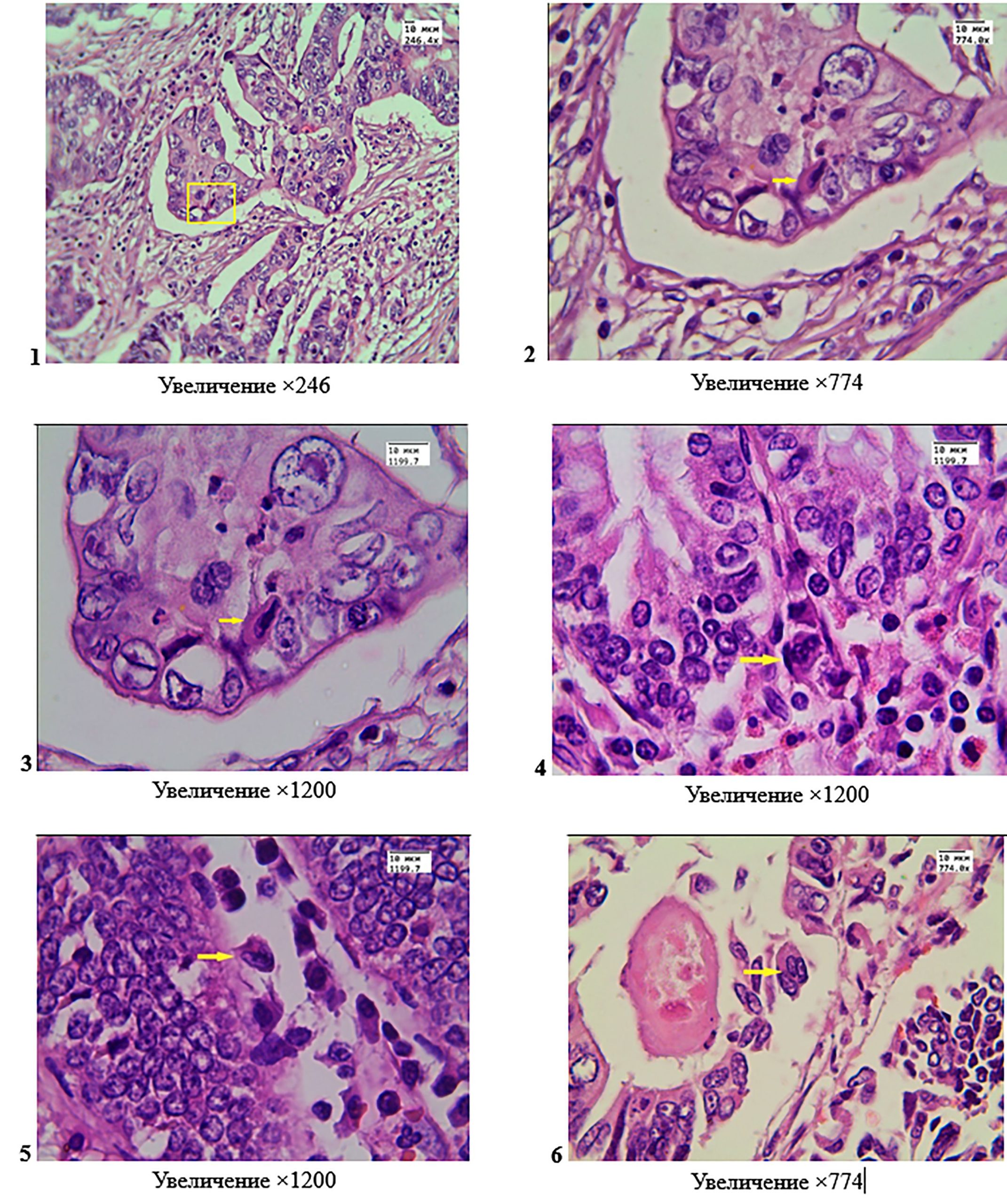
Surgical preparations of resected colon segments for tumors with the structure of moderately differentiated adenocarcinoma, hematoxylin and eosin staining. Photos 1, 2, 3 – a binuclear cell (shown by a square and arrows) with two phenotypically different nuclei, one of which is hyperchromatic; the cell has morphological features of a heterokaryon of hybrid origin; the cell is located among the epithelial cells of the tumor tissue. Photos 4, 5, 6 – among the crypts there are multinuclear cells (shown by arrows), the nuclei of which are phenotypically similar to the nuclei of epithelial cells; the cells are not associated with the basal membrane; photo 6 – a cell in which 3 nuclei are clearly visible due to the fusion of epithelial cells, which indicates its hybrid origin.